The US National Institute of Allergy and Infectious Diseases released photos of SARS-CoV-2, the pathogen responsible for covid-19 infection
The U.S. National Institute of Allergy and Infectious Diseases (NIAID) released a new series of images of the SARS-CoV-2 virus, which is the pathogen responsible for covid-19 infection.
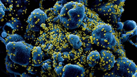
In the photographs, diffused through the Flickr platform, you can see how these microorganisms, with an appearance similar to small spheres, surround the cells of a patient infected with the disease.
As detailed, the cells are in the process of apoptosis, or self-destruction.

How the size of the virus is around 100 nanometers, well below what a traditional lens can perceive, the images were captured with an electron microscope and later colored.

Like the other coronaviruses, SARS-CoV-2 is basically a ball of ribonucleic acid (RNA), which contains its genetic information. Its lipid surface is endowed with various protein ‘spines’, which allow it to adhere to the cell and force it to replicate the viral RNA as if it were its own.
Earlier this month, Chinese scientists disclosed the virus’s actual appearance, also photographing it with an electron microscope.
Find out the latest on digital economy, startups, fintech, corporate innovation and blockchain. CLICK HERE

Corresponsal de Argentina, Encargado de seleccionar las noticias más relevantes de su interés a nuestro sitio web NewsPer.com

